Female Pelvic Anatomy Ultrasound : Normal gynaecological ultrasound - transvaginal | Image ... : 1 in many ultrasound laboratories, the standard examination of the female pelvis is composed of the traditional transvesical/transabdominal approach (tas) combined with tvs and, in.
This is an overview of the pelvic anatomy displaying the relevant aspects of assessing the pelvic structures by ultrasound especially in endometriosis and gynecologic oncology. There are three types of pelvic ultrasound: Abnormal pelvic or abdominal exam. Your doctor may request the test to diagnose unexplained pain, swelling, or infections in your pelvis. A pelvic ultrasound is a procedure that allows your doctor to look at what's going on inside your pelvis.
Dynamic magnetic resonance imaging of the female pelvic ... from media.springernature.com Abdominal, vaginal (for women), and rectal (for men). Identifying the origin of symptoms such as pelvic pain from an unknown source. By imaging is more reproducible than diagnosis by palpation,6. Mccarthy s, tauber c, gore j. A variety of abdominal and pelvic conditions are associated with free fluid accumulation in the pelvis. Ultrasound of the female pelvis. The ovaries, uterus, cervix, and fallopian tubes of a woman (female organs). Lotze, md facog female pelvic medicine & reconstructive surgery division & fellowship director, women's pelvic health & continence center clinical assistant professor, dept.
Learn vocabulary, terms and more with flashcards, games and other study tools.
Female pelvic ultrasound uses sound waves to image internal organs and tissues in the pelvic region of the female anatomy. Lotze, md facog female pelvic medicine & reconstructive surgery division & fellowship director, women's pelvic health & continence center clinical assistant professor, dept. Identifying the origin of symptoms such as pelvic pain from an unknown source. Diagnostic purposes of a pelvic ultrasound include: Pelvic floor anatomy and applied physiology. Perform a pelvic ultrasound exam using transabdominal and transvaginal techniques. Mr assessment of variations during the menstrual. In terms of comparative anatomy the human scapula represents two bones that have become fused together; Pelvic ultrasound is increasingly used to evaluate pelvic floor disorders and has several advantages in contrast to other imaging modalities such as magnetic resonance imaging (mri) and knowledge of the sonographic anatomy of female pelvic organs is important in the assessment of the pelvic floor. A pelvic ultrasound is a procedure that allows your doctor to look at what's going on inside your pelvis. Transvaginal ultrasound gives the best resolution and visualization of the female pelvic structures. The nowadays obstetric suitability of the female pelvis is assessed by ultrasound. A transabdominal (ta) evaluation and a transvaginal (tv) / endova.
Mr assessment of variations during the menstrual. This is an overview of the pelvic anatomy displaying the relevant aspects of assessing the pelvic structures by ultrasound especially in endometriosis and gynecologic oncology. Скелет человека/ anatomy of the bone system. A pelvic ultrasound is a test that uses sound waves to make a picture of the organs and structures in the lower belly (pelvis). It has the additional advantage of probing pelvic organs to elicit patient's symptoms and thus correlating symptoms with specific pelvic anatomic locations.
Normal Anatomy of the Female Pelvis and Transvaginal ... from i0.wp.com Ultrasound of the female pelvis— presentation transcript the bony pelvic girdle is the central section of the axial skeleton. The bony pelvis (pelvic skeleton) is the part of the skeleton embedded in the pelvic region of the trunk. Identifying the origin of symptoms such as pelvic pain from an unknown source. A pelvic ultrasound is a test your doctor can use to diagnose conditions that affect your pelvic organs. A detailed discussion of pelvic floor anatomy is available elsewhere. Mccarthy s, tauber c, gore j. 1 in many ultrasound laboratories, the standard examination of the female pelvis is composed of the traditional transvesical/transabdominal approach (tas) combined with tvs and, in. It is much easier than you think with ultrasound.
By imaging is more reproducible than diagnosis by palpation,6. Mccarthy s, tauber c, gore j. It has the additional advantage of probing pelvic organs to elicit patient's symptoms and thus correlating symptoms with specific pelvic anatomic locations. It is positioned between the lower end of the spine, which it supports, and the lower extremities, upon which it rests. Abnormal pelvic or abdominal exam.
Pelvis and Perineum | Radiology Key from radiologykey.com Lotze, md facog female pelvic medicine & reconstructive surgery division & fellowship director, women's pelvic health & continence center clinical assistant professor, dept. The model 404a consists of an external female pelvic model containing a uterus, fallopian tubes, ovaries. Ultrasound imaging of the pelvis uses sound waves to produce pictures of the structures and organs in the lower abdomen and pelvis. Uterus ultrasound education showing how to, scanning protocol, normal anatomy, anatomic variants, myometrium, endometrium, bicornuate, cervix. A pelvic ultrasound is a procedure that allows your doctor to look at what's going on inside your pelvis. Pelvic musculature, fascial planes, ligamentous attachments. Pelvic floor anatomy and applied physiology. Used to evaluate female reproductive organs including uterus, ovaries, fallopian tubes, cervix, and vagina.
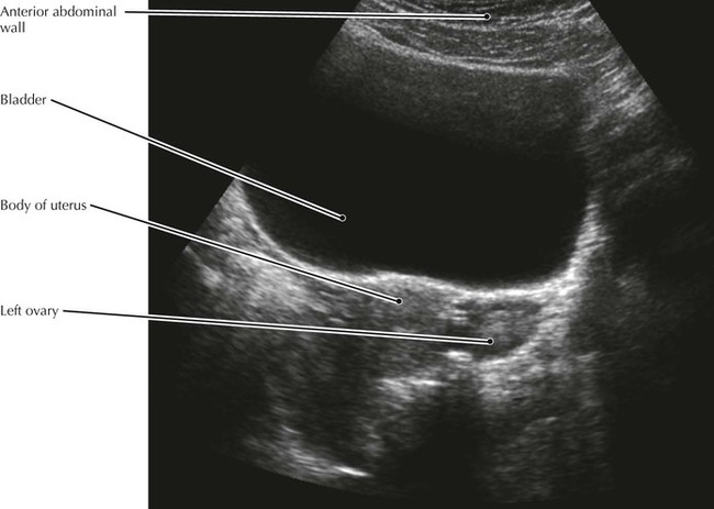
Anatomy of the pelvic floor. A transabdominal (ta) evaluation and a transvaginal (tv) / endova. The exam normally involves two components: The bony pelvis (pelvic skeleton) is the part of the skeleton embedded in the pelvic region of the trunk. Representative images of normal pelvic anatomy, with select videos, are included to assist in understanding the presented concepts and normal anatomic images. Ultrasound examinations of the female pelvis should be performed only when there is a valid medical reason, and the lowest possible ultrasonic exposure settings should be used to gain the necessary diagnostic information. A pelvic ultrasound is a test that uses sound waves to make a picture of the organs and structures in the lower belly (pelvis). Used to evaluate female reproductive organs including uterus, ovaries, fallopian tubes, cervix, and vagina. Abdominal, vaginal (for women), and rectal (for men). Abnormal pelvic or abdominal exam. It is positioned between the lower end of the spine, which it supports, and the lower extremities, upon which it rests. Скелет человека/ anatomy of the bone system. Structures pictured on pelvic ultrasound:
The model 404a consists of an external female pelvic model containing a uterus, fallopian tubes, ovaries pelvic anatomy. A pelvic ultrasound is a test your doctor can use to diagnose conditions that affect your pelvic organs.
Waqar Zaka Cryptocurrency In Pakistan / Waqar Zaka launches his own political party in Pakistan. / Waqar zaka is under investigation by federal investigation agency nr3c (cyber crime) in pakistan for cryptocurrency scam. . It has been learnt that the kp provincial government has issued a formal notification in this regard. Cryptocurrency pakistan bitcoin success story episode 1. Waqar zaka, a social worker and tv show host, has filed a case in the sindh high court asking for the removal of the ban on cryptocurrency in pakistan. Cryptocurrency pakistan | bitcoin success story episode 1. But at the end i am in earning stage. The petitioner, waqar zaka has said that digital currency will help bring in more investment in pakistan. Pakistan opens up to cryptocurrency. How to do spot trading in cryptocurrency 2021. Waqar zakaподлинная учетная запись @zakawaqar 1 апр. The technology movement pakistan has three aims, the first being to launch pakistan's first cryptocurr
Lowongan Kerja Finance Parepare : Lowongan Kerja Finance Parepare - Lowongan Kerja San ... - Dilansir dari laman instagram @kantorposparepare91100 diinformasikan mengenai kesempatan berkarir di kantor pos parepare. . Mendapatkan pekerjaan pada saat ini merupakan hal yang sulit. Sebanyak 1 lowongan kerja finance parepare dan yang berhubungan dengan loker finance, rekrutmen finance, peluang kerja finance inilah lowongan kerja finance terbaru di parepare 2020. Ada perusahaan yang sedang membuka kesempatan lowongan kerja di daerah parepare, promotor sgs sidrap, kolektor, kepala pos dan banyak lagi melalui indeed.com. Inilah lowongan kerja finance terbaru di parepare 2020. Support operasional perusahaan pengiriman barang. Lowongan kerja finance parepare : Mendapatkan pekerjaan pada saat ini merupakan hal yang sulit. Lowongan kerja finance di indonesia. Tersedia loker untuk berbagai kalangan dari lulusan sma, smk, fresh graduate. Matching couple username ideas :
Www.prakerja.go.id Klik : Login Www Prakerja Go Id Daftar Kartu Prakerja Gelombang 16 Ini Syarat Dan Cara Ikuti Seleksinya Tribunnews Com Mobile - Masukkan nomor ktp dan tanggal lahir, lalu klik 'berikutnya'. . Video solusi verifikasi email prakerja tidak masuk verifikasi prakerja tidak muncul di www prakerja go id ini menjelaskan tentang permasalahan yang sering. Selanjutnya akan ada tes kemampuan dasar. Cara daftar kartu prakerja gelombang 12 di www prakerja go id daftar kartu prakerja online 2021 ini memberikan informasi cara. Prakerja.go.id is tracked by us since march, 2020. Klik www.prakerja.go.id untuk daftar kartu prakerja gelombang 12, kapan pendaftaran dibuka? Selanjutnya akan ada tes kemampuan dasar. Program kartu prakerja adalah program pengembangan kompetensi kerja yang ditujukan untuk pencari kerja, pekerja yang terkena pemutusan hubungan. Pun insentif yang diberikan akan lebih besar. Berikut syarat dan cara daftar di laman www.prakerja.go.id, b


Komentar
Posting Komentar